What is the most posterior structure of the Atlas C1 vertebra?
Similarly, it is asked, what is posterior to the atlas?
Gross anatomy
The transverse ligament holds the dens of the axis against the anterior arch of the atlas and divides its vertebral canal into two parts. The anterior 1/3 is occupied by the dens. The posterior 2/3 contains the spinal cord, which occupies 1/3 of the total vertebral canal space.
One may also ask, what is unique about C1 vertebra? Anatomy and function
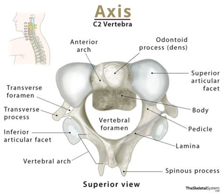
The first cervical vertebra, atlas, is unique in the sense that it possesses neither a body nor spinous process. It consists of an anterior and posterior arch extending between two lateral masses, forming a closed triangular 'ring' that accommodates the brainstem.
Thereof, how we can distinguish anterior and posterior arches of Atlas?
The anterior arch contains a facet for articulation with the dens of the axis. This is secured by the transverse ligament of the atlas - which attaches to the lateral masses. The posterior arch has a groove for the vertebral artery and C1 spinal nerve.
Is C1 same as Atlas?
In anatomy, the atlas (C1) is the most superior (first) cervical vertebra of the spine and is located in the neck. It is named for Atlas of Greek mythology because, just as Atlas supported the globe, it supports the entire head.
| Atlas (anatomy) | |
|---|---|
| TA98 | A02.2.02.101 |
| TA2 | 1038 |
| FMA | 12519 |
| Anatomical terms of bone | |
Related Question Answers
Can you adjust your own Atlas?
With our exercises' help, you can usually make an atlas correction yourself by loosening and stretching the muscles and fasciae in this area. An atlas blockage can also be located between the atlas vertebra and the underlying axis.What is the Atlas in the human body?
The Atlas: The Top Bone in Your Cervical SpineThe occipital bone rests upon the atlas, the first bone in your neck. The atlas is named after the Greek God Atlas, who held up the world on his shoulders. A pair of synovial joints, known as the atlanto-occipital joint connect the atlas and your skull.
Why is it called the atlas bone?
The atlas bone is the first of seven cervical vertebrae (vertebra cervicalis I or C1). It supports the weight of the skull. The name for the bone was derived from a deity of Greek mythology called Atlas, who supported the heavens.Where is the posterior arch of Atlas?
Description. The posterior arch forms about two-fifths of the circumference of the ring: it ends behind in the posterior tubercle, which is the rudiment of a spinous process and gives origin to the Recti capitis posteriores minores.What does the first cervical vertebra C1 or Atlas lack?
Assoc Prof Craig Hacking ? ? and Dr Dinesh Palipana et al. Of the cervical vertebrae, the atlas (C1), axis (C2) and vertebra prominens (C7) are considered atypical cervical vertebrae. The atlas (C1) lacks a body or spinous process. It has anterior and posterior arches with lateral masses.Why is C2 called axis?
C2 (cervical vertebra): C2 is the symbol for the second cervical vertebra, which is also called the axis. It is so-named because the uppermost cervical vertebra (called the atlas) rotates about the odontoid process of the second cervical vertebra.Does Atlas have Pedicles?
The atlas is made up of a thick anterior arch, a thin posterior arch, 2 prominent lateral masses, and 2 transverse processes. The axis is composed of a vertebral body, heavy pedicles, laminae, and transverse processes, which serve as attachment points for muscles.What is the difference between Atlas and Axis vertebrae?
The atlas is the first cervical (neck) vertebra which is just under the head; it is named for Atlas, the Greek god who supported the world on his shoulders. The axis is the second cervical vertebra; it has what is called the odontoid process about which the atlas rotates. It allows the head turn from side to side.Which joint is present in neck?
Pivot joints, such as the neck joints, allow limited rotating movements.Why is the neck called cervical?
The word cervix is derived from the Latin root word "cervix" which means "neck." For this reason, the word cervical pertains to many areas where tissues narrow to a neck-like passage, and not only in your neck.Does C1 have a vertebral body?
C1. The Atlas, C1, is the topmost vertebra, and along with the Axis; forms the joint connecting the skull and spine. Its chief peculiarity is that it has no body, and this is due to the fact that the body of the atlas has fused with that of the Axis.How many cervical vertebrae are in the human body?
The spine above the sacrum consists of: Seven bones in the neck—the cervical spine. 12 bones in the chest—the thoracic spine. Five bones in the lower back—the lumbar spine.What is unique about the hyoid bone?
The hyoid bone is a unique structure in the human body for many reasons. Famously, the hyoid bone is the only bone in humans that does not articulate with any other bone, but only has muscular, ligamentous, and cartilaginous attachments. Given this peculiarity, it has been described as “free floating” [1].How do you palpate Atlas?
The transverse process of the atlas (C1) can be palpated inferior to the ear between the angle of the mandible and the styloid process of the temporal bone. On the lateral aspect of the skull, about 4 cm superior to the midpoint of the zygomatic arch, is the pterion.How do I adjust my atlas bone?
To adjust your atlas, we use an Atlas Orthogonal Precision Adjusting Instrument. This instrument may sound intimidating, but it's actually completely painless - literally, you won't feel a thing. You simply lie on your side and the doctor places the tip of the instrument just below your earlobe.What are the 7 cervical vertebrae?
Position of human cervical vertebrae (shown in red). It consists of 7 bones, from top to bottom, C1, C2, C3, C4, C5, C6, and C7.Why are C1 and C2 special?
The C1 vertebrae is named atlas and the C2 vertebrae is named axis. As well as protecting the spinal cord, these vertebrae are primarily responsible for facilitating and controlling the large range of movement that your neck has, and supporting the considerable weight of your skull at the tip of your spine.What nerves are affected by C1 and C2?
C1, C2, and C3 (the first three cervical nerves) help control the head and neck, including movements forward, backward, and to the sides. The C2 dermatome handles sensation for the upper part of the head, and the C3 dermatome covers the side of the face and back of the head. (C1 does not have a dermatome.)What happens when you break your C1 vertebra?
There may be pain and stiffness, usually isolated to the area around the fractured vertebra. You may have trouble walking and even breathing if there's been spinal cord damage. You may feel a lot of pain in another part of the body and not be aware of your neck pain.What nerves are affected by C1?
The cervical spinal nerve 1 (C1) is a spinal nerve of the cervical segment. C1 carries predominantly motor fibres, but also a small meningeal branch that supplies sensation to parts of the dura around the foramen magnum (via dorsal rami).Cervical spinal nerve 1.
| Cervical spinal nerve | |
|---|---|
| Latin | Nervi spinalis |
| FMA | 6440 |
| Anatomical terminology | |
What causes C1 misalignment?
A misalignment at C0-C1-C2 can also come from sitting at a desk with poor posture, birth trauma, or falling out of a tree when you are a child. However, the most common way we see large trauma produced is from a concussion from either sports or work related injuries, or a car accident.Can C1 and C2 vertebrae misalignment symptoms?
The atlanto-axial joint allows for healthy rotation in your neck, but it can cause the C1 and C2 to become misaligned when it's affected by RA. Misalignment and instability can impinge your spinal cord, brainstem and/or other nerves, which can cause pain and other serious neurological symptoms.What is cervical vertebrae 1 C1 called?
The C1 and C2 vertebrae are the first two vertebrae of the cervical spine. They are also called the atlas and axis vertebrae.Can C1 and C2 cause headaches?
Role of Spinal NervesC1, C2 and/or C3 may be involved in development of cervicogenic headaches because these nerves enable function (movement) and sensation of the head and neck. Nerve compression can cause inflammation and pain.
What is C1 C2 subluxation?
(C1–C2 Subluxation)Atlantoaxial subluxation is misalignment of the 1st and 2nd cervical vertebrae, which may occur only with neck flexion. (See also Evaluation of Neck and Back Pain and Craniocervical Junction Abnormalities.)