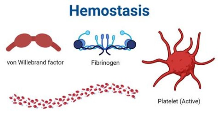
Hemostasis is the mechanism that leads to cessation of bleeding from a blood vessel. It is a process that involves multiple interlinked steps. This cascade culminates into the formation of a “plug†that closes up the damaged site of the blood vessel controlling the bleeding. Also to know is, what is the function of hemostasis?
Hemostasis is derived from a Greek word, which means stoppage of blood flow. The process is a combination of cel- lular and biochemical events that function together to keep blood in the liquid state within the veins and arteries and prevent blood loss following injury through the formation of a blood clot.
Similarly, how many systems does hemostasis have? The hemostatic system comprises platelet aggregation, coagulation and fibrinolysis also termed primary, secondary and tertiary hemostasis.
Also Know, what is hemostasis and why is it important?
Hemostasis or haemostasis is a process to prevent and stop bleeding, meaning to keep blood within a damaged blood vessel (the opposite of hemostasis is hemorrhage). It is the first stage of wound healing. This involves coagulation, blood changing from a liquid to a gel.
What is normal hemostasis?
Normal hemostasis is best conceptualized according to its major components—vessel wall, platelet function, coagulation factor cascade, and clot inhibition/fibrinolysis. These components work together to prevent prolonged bleeding or thrombosis under normal physiologic conditions.
Related Question Answers
What are the 4 steps of hemostasis?
Hemostasis includes three steps that occur in a rapid sequence: (1) vascular spasm, or vasoconstriction, a brief and intense contraction of blood vessels; (2) formation of a platelet plug; and (3) blood clotting or coagulation, which reinforces the platelet plug with fibrin mesh that acts as a glue to hold the clot What are the two major disorders of hemostasis?
The most common inherited diseases are von Willebrand disease (primary hemostasis), which is the most common inherited disorder of hemostasis, and hemophilia A (factor VIII deficiency, secondary hemostasis). How do you get hemostasis?
A variety of hemostatic methods can be employed, ranging from simple manual pressure application with one finger to electrical tissue cauterization, systemic administration of blood products, and systemic administration or topical application of procoagulation agents. What are the factors that affect hemostasis?
It is affected by the characteristics of blood vessel walls, platelets, the fibrinolytic system, and the coagulation pathway, which are all intimately related ( Figure 1). All these factors function normally to produce an equilibrium between antithrombotic and prothrombotic factors. How does primary hemostasis work?
Primary hemostasis serves to immediately limit bleeding through the formation of a loose platelet plug. Platelets play a key role in the rapid response to blood vessel injury by: Adhering to the endothelial wall at the site of injury. Releasing potent anticoagulant compounds. What occurs in primary hemostasis?
Primary hemostasis refers to platelet aggregation and platelet plug formation. Platelets are activated in a multifaceted process (see below), and as a result they adhere to the site of injury and to each other, plugging the injury. What is the difference between primary and secondary hemostasis?
Primary hemostasis is a procoagulation clot forming process associated with the initiation and formation of the platelet plug. Secondary hemostasis also a procoagulation clot forming process and it is associated with the propagation of the clotting process via the intrinsic and extrinsic coagulation cascades. Why is secondary hemostasis necessary?
Secondary hemostasis is triggered by the release of tissue factor from epithelial cells that are exposed to the circulation at the site of vascular injury. Defects in secondary hemostasis decrease fibrin production and reduce the stability of the formed clot. What activates platelets during hemostasis?
Platelets contain secretory granules. When they stick to the proteins in the vessel walls, they degranulate, thus releasing their products, which include ADP (adenosine diphosphate), serotonin, and thromboxane A2 (which activates other platelets). Which blood cells are called thrombocytes?
The main job of platelets, or thrombocytes, is blood clotting. Platelets are much smaller in size than the other blood cells. They group together to form clumps, or a plug, in the hole of a vessel to stop bleeding. What causes fibrinolysis?
Primary fibrinolysis occurs naturally and secondary fibrinolysis occurs due to an external cause such as medicine or a medical disorder. Fibrinolysis is tightly controlled by the actions of various cofactors, inhibitors, and receptors. Plasmin is the main protein that activates fibrinolysis. What do platelets do?
Platelets, or thrombocytes, are small, colorless cell fragments in our blood that form clots and stop or prevent bleeding. Platelets are made in our bone marrow, the sponge-like tissue inside our bones. What's the meaning of fibrinogen?
: a plasma protein that is produced in the liver and is converted into fibrin during blood clot formation. Why does blood clot happen?
Blood clots form when certain parts of your blood thicken, forming a semisolid mass. This process may be triggered by an injury or it can sometimes occur inside blood vessels that don't have an obvious injury. How does hemostasis affect platelets?
Released secretary granules will recruit additional platelets to form the platelet plug, which is referred to as primary hemostasis10. Following vasoconstriction, exposed collagen from the damaged surface will encourage platelets to adhere, activate and aggregate to form a platelet plug, sealing off the injured area. What does embolus mean?
Embolus: A blockage or plug that obstructs a blood 'vessel. Examples of emboli are detached blood clots, clumps of bacteria, and clumps of other foreign material, such as air. What is clot retraction called?
Clot retraction generally occurs within 24 hours of initial clot formation and decreases the size of the clot by 90%. Following clot retraction, a separate process called fibrinolysis occurs which degrades the fibrin of the clot while macrophages consume the expended platelets, thus preventing possible thromboembolism. Can you dissolve blood clots naturally?
Typically, your body will naturally dissolve the blood clot after the injury has healed. Sometimes, however, clots form on the inside of vessels without an obvious injury or do not dissolve naturally. These situations can be dangerous and require accurate diagnosis and appropriate treatment. What is hemostasis testing?
The screening tests of hemostasis were developed to help identify patients with hemostatic defects that could cause excessive bleeding. Screening tests are available for each of the three phases of hemostasis: coagulation (fibrin clot formation), platelet plug formation, and fibrinolysis. What is hemostasis in hematology?
Hemostasis, the arrest of bleeding from an injured blood vessel, requires the combined activity of. Vascular factors. Platelets. Plasma coagulation factors. What is hemostasis PDF?
Hemostasis governs two essential processes of human life in that it maintains the fluidity of blood under physiological conditions and prevents excessive blood loss after injury. Hemostasis is regulated by components of the vessel wall and blood cells and by humoral coagulation factors. What is mechanism of blood clotting?
The mechanism of coagulation involves activation, adhesion, and aggregation of platelets along with deposition and maturation of fibrin. Disorders of coagulation can result in bleeding (hemorrhage or bruising) or obstructive clotting (thrombosis). What is thrombosis of a vessel?
Thrombosis occurs when blood clots block your blood vessels. There are 2 main types of thrombosis: Venous thrombosis is when the blood clot blocks a vein. Veins carry blood from the body back into the heart. Arterial thrombosis is when the blood clot blocks an artery. How do you measure bleeding time?
A standard-sized incision is made around 10 mm long and 1 mm deep. The time from when the incision is made until all bleeding has stopped is measured and is called the bleeding time. Every 30 seconds, filter paper or a paper towel is used to draw off the blood. The test is finished when bleeding has stopped.