What is a t1 and t2 hyperintense lesion?
Keeping this in view, what is hyperintense on t1 and t2?
Hyperintensity is a term used in MRI reports to describe how part of an image looks on MRI scan. There are a variety of MRI sequences or imaging patterns used (ie. T1, T2 or FLAIR) to highlight or suppress different types of tissue so that abnormalities can be detected.
Similarly, what are t1 and t2 lesions? Specifically, T1 and T2 refers to the time taken between magnetic pulses and the image is taken. These different methods are used to detect different structures or chemicals in the central nervous system. T1 and T2 lesions refers to whether the lesions were detected using either the T1 or T2 method.
Also Know, what is a t1 hyperintense lesion?
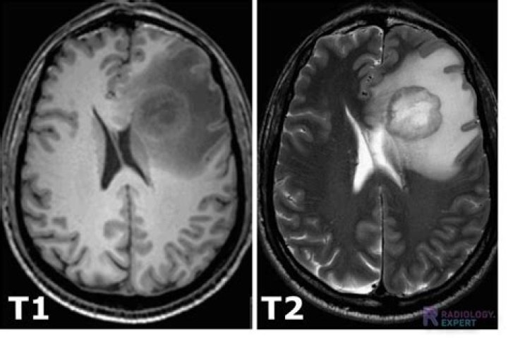
Abstract. T1 -hypointense lesions (T1-black holes) in multiple sclerosis (MS) are areas of relatively severe central nervous system (CNS) damage compared with the more non-specific T2-hyperintense lesions, which show greater signal intensity than normal brain on T2-weighted magnetic resonance imaging (MRI).
What is a t2 hyperintense lesion?
White matter hyperintensities (WMHs) are lesions in the brain that show up as areas of increased brightness when visualised by T2-weighted magnetic resonance imaging (MRI). WMH's are also referred to as Leukoaraiosis and are often found in CT or MRI's of older patients.
Related Question Answers
What does t2 mean in medical terms?
T2 in Medical| T2 | Type 2 Diabetic Infertility |
|---|---|
| T2 | spin-spin or transverse relaxation time Medicine, Healthcare, Science |
| T2 | Type 2 Diabetic - Insulin Resistant, Adult Onset Infertility, Health |
| T2 | breast tumor 2.0 to 5.0 cm Medicine, Healthcare, Science |
| T2 | diiodothyronine Medicine, Healthcare, Science |
What is t1 and t2 in the brain?
The basic types of sequences used in brain MRI create either T1-weighted or T2-weighted images. In T1-weighted images, CSF and fluid appear dark. In T2-weighted images, CSF and fluid have a higher signal intensity than tissue and therefore appear bright.What causes t2 hyperintensity?
Causes. White matter hyperintensities can be caused by a variety of factors including ischemia, micro-hemorrhages, gliosis, damage to small blood vessel walls, breaches of the barrier between the cerebrospinal fluid and the brain, or loss and deformation of the myelin sheath.What is the difference between hypointense and hyperintense?
Often we refer to the appearance by relative terms: hyperintense = brighter than the thing we are comparing it to. isointense = same brightness as the thing we are comparing it to. hypointense = darker than the thing we are comparing it to.What is the relationship between t1 and t2?
Hence solids, macromolecules, and bound water molecules rotate slowly and have short T2 values. T2 progressively increases with molecular tumbling rate. In the limit (pure liquids like CSF) T1=T2 and both are several seconds in length. The overall T1 effect can be thought of as a "Goldilock's" phenomenon.What is t2 and flair Hyperintensities?
Focal hyperintensities in the subcortical white matter demonstrated by T2-weighted or FLAIR images are a common incidental finding in patients undergoing brain MRI for indications other than stroke. They are indicative of chronic microvascular disease. These white matter lesions may progress in number and frequency.What is a t2 brain lesion?
T2/FLAIR lesions can directly account for some symptoms. For example, a brainstem lesion can cause room spinning sensations and balance problems. Cervical (neck) spinal cord T2/FLAIR lesions could cause tingling and numbness in the hands and legs. Many of the lesions may not be causing obvious symptoms.What does increased t1 signal mean?
T1 weighted image – Pathology (spine)Loss of the normal high signal in the bone marrow indicates loss of normal fatty tissue and increased water content. Abnormal low signal on T1 images frequently indicates a pathological process such as trauma, infection, or cancer.
What appears bright on t1?
T1 weighting tends to have short TE and TR times. Fat quickly realigns its longitudinal magnetization with B0, and it therefore appears bright on a T1 weighted image.What does t1 and t2 mean in MRI?
The most common MRI sequences are T1-weighted and T2-weighted scans. T1-weighted images are produced by using short TE and TR times. The contrast and brightness of the image are predominately determined by T1 properties of tissue. Conversely, T2-weighted images are produced by using longer TE and TR times.What does hypointense signal mean?
Hypointense signal changes on T2-weighted images were defined as areas of signal intensity equal or lower to signal intensity of the globus pallidus according to prior studies on putaminal hypointensities in parkinsonism.What is stir hyperintense lesion?
Vertebral hemangiomas are typically well-circumscribed, benign vascular tumors, which are T1 hyperintense (Figure 15). These lesions may be dark or bright on STIR sequences dependent on the proportion of fatty and vascular elements. The coarse vertical trabeculae resemble corduroy or honeycomb of radiographs.What does increased Flair signal mean?
FLAIR MRI is a heavily T2-weighted technique that dampens the ventricular (ie, free-water) CSF signal. Thus, the highest signals on the sequence are from certain brain parenchymal abnormalities, such as MS lesions, while the CSF appears black.What is a hyperintense lesion on spine?
T1 hyperintense bone lesions are virtually always benign. However, correlation with the lesion appearances on other MR imaging sequences and imaging modalities as well as with the clinical history may occasionally suggest otherwise. The vast majority of T1 hyperintense vertebral column lesions are benign.What is t2 hyperintensity in spinal cord?
Hyperintense intramedullary signal at T2-weighted imaging is a common and important indicator of myelopathy at MRI (1). T2 hyperintensity can reflect many processes at the microscopic level, including edema, blood–spinal cord barrier breakdown, ischemia, myelomalacia, or cavitation (2).What is increased t2 signal on MRI report?
An increase in T2 signal intensity is often associated with chronic compression of the spinal cord, and it is well established that chronic compression results in structural changes to the spinal cord.How do you remember t1 vs t2 MRI?
Radiology MnemonicHere's an easy way to remember MRI image weighting using Arnold Schwarzenegger and the Terminator Movies: Just use the Terminator movies to remember what water will look like on a T1 or T2 wieghted MRI!!!!
What color is blood on MRI?
The center of chronic hematomas usually have high water content, rendering them bright, not dark, on T2-weighted images. The periphery of chronic hematomas contain hemosiderin, rendering them slightly dark on T2-weighted images but profoundly dark on T2*/SW images.Does white matter lesions mean MS?
White matter tracts are affected, including those of the cerebral hemispheres, infratentorium, and spinal cord. MS lesions, known as plaques, may form in CNS white matter in any location; thus, clinical presentations may be diverse.What does t1 flair mean?
T1-FLAIR stands for T1-weighted-Fluid-Attenuated Inversion Recovery. This nomenclature began to arise in the late 1990s to denote an inversion recovery sequence with dark CSF and other T1-like properties made possible by a medium TI coupled with fast spin-echo signal acquisition.How many lesions is alot for MS?
An “average” number of lesions on the initial brain MRI is between 10 and 15. However, even a few lesions are considered significant because even this small number of spots allows us to predict a diagnosis of MS and start treatment. Q2.Are white spots on brain MRI normal?
The finding of a "white matter lesion" in the brain during an MRI is quite common. Its significance depends on the patient's presentation.Are white matter lesions serious?
Serious consequences of periventricular white matter lesions -- this is the scary part. There is strong evidence that cerebral white matter lesions impair brain function, and in particular impair thinking ability and walking.Can white matter lesions in the brain be nothing?
White matter lesions observed on brain MRI are usually characteristic and occur in specific areas including the corpus callosum and pons. “However, in many cases, the white matter lesions as isolated observations are nonspecific” and could be due to MS or another cause, explained Drs Lange and Melisaratos.Can migraines cause white spots on brain MRI?
In addition, multiple studies have found that people with migraines have an increased risk of brain lesions. The two main types of lesions found in migraineurs include: White matter hyperintensities (WMH): These lesions appear bright white on certain sequences of MRI scans.What diseases cause white matter on the brain?
White matter disease is a disease that affects the nerves that link various parts of the brain to each other and to the spinal cord.Risk factors for white matter disease may include:
- smoking cigarettes.
- older age.
- heart disease.
- high blood pressure.
- high cholesterol.