What does a PE look like on EKG?
Beside this, does pulmonary embolism show up on EKG?
Chest X-ray is often normal in pulmonary embolism. EKG may be normal, but may also show indirect signs of PE. These include tachycardia (heart rate >100), and changes associated with right ventricle strain.
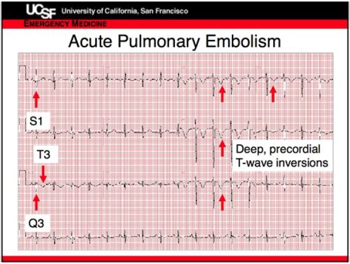
Furthermore, what does s1q3t3 mean? S1Q3T3 pattern was defined as the presence of S wave in lead I and Q wave and inverted T wave in lead III. Prior cardiopulmonary disease was defined as a prior diagnosis or evidence of chronic cardiac or pulmonary diseases.
Secondly, can an EKG tell if you have a blood clot?
Other tests: An X-ray or ECG / EKG is not normally a test which will be recommended for the diagnosis of a blood clot, but may be requested if there are signs of other concerns relating to certain symptoms.
What does pulmonary disease pattern mean on ECG?
ECG Examples
ECG demonstrates many of the features of chronic pulmonary disease: Rightward QRS axis (+90 degrees). Peaked P waves in the inferior leads > 2.5 mm (P pulmonale) with a rightward P-wave axis (inverted in aVL) Clockwise rotation of the heart with a delayed R/S transition point (transitional lead = V5).
Related Question Answers
What's the most common ECG finding in a patient with a pulmonary embolism?
The most common ECG finding in the setting of a pulmonary embolism is sinus tachycardia. However, the “S1Q3T3” pattern of acute cor pulmonale is classic; this is termed the McGinn-White Sign. A large S wave in lead I, a Q wave in lead III and an inverted T wave in lead III together indicate acute right heart strain.What does a blood clot in your lung feel like?
The feeling can range from a dull ache to intense pain. Trouble breathing. If this happens, it could mean that the clot has moved from your arm or leg to your lungs. You may also get a bad cough, and might even cough up blood.Can you have a PE and not know it?
Although most people with a pulmonary embolism experience symptoms, some will not. The first signs are usually shortness of breath and chest pains that get worse if you exert yourself. You may cough up bloody sputum. If you have these symptoms get medical attention right away.How long can a pulmonary embolism go undetected?
Symptoms from a pulmonary embolism, like shortness of breath or mild pain or pressure in your chest, can linger 6 weeks or more. You might notice them when you're active or even when you take a deep breath. Exercise can help with this.Is elevated D dimer serious?
If your results show higher than normal levels of D-dimer, it may mean you have a clotting disorder. But it cannot show where the clot is located or what type of clotting disorder you have. Also, high D-dimer levels are not always caused by clotting problems.Can a CT scan detect a blood clot in the lungs?
CT pulmonary angiography ? also called CT pulmonary embolism study ? creates 3D images that can detect abnormalities such as pulmonary embolism within the arteries in your lungs. In some cases, contrast material is given intravenously during the CT scan to outline the pulmonary arteries.How long can you live with blood clots in your lungs?
Medium to long term. After the high-risk period has elapsed (roughly one week), blood clots in your lung will need months or years to completely resolve. You may develop pulmonary hypertension with life-long implications, including shortness of breath and exercise intolerance.Can a blood test detect pulmonary embolism?
Your doctor will order a D-dimer blood test to help diagnose or rule out the presence of a pulmonary embolism. The D-dimer test measures the levels of a substance that is produced in your bloodstream when a blood clot breaks down.Can urgent care check for a blood clot?
If you notice these symptoms, go to your nearest urgent care. They can usually diagnose a DVT with a simple ultrasound and give you medications to help resolve the blockage. DVT is dangerous because if more clots form or if the clot moves from your leg, it could cause a heart attack, pulmonary embolism, or a stroke.Can you have a pulmonary embolism for months without knowing?
DVT often goes undetected, because symptoms, such as pain or swelling in the leg, shortness of breath, chest pain, coughing and dizziness, are missed or dismissed as minor. And in some cases, there are no symptoms until it is too late.Can EKG detect a blockage?
Your doctor may use an electrocardiogram to determine or detect: Abnormal heart rhythm (arrhythmias) If blocked or narrowed arteries in your heart (coronary artery disease) are causing chest pain or a heart attack. Whether you have had a previous heart attack.How do you know if you have a Bloodclot?
Symptoms of a blood clot include:- throbbing or cramping pain, swelling, redness and warmth in a leg or arm.
- sudden breathlessness, sharp chest pain (may be worse when you breathe in) and a cough or coughing up blood.
Can Xrays show clots?
This test uses X-rays to show your deep veins. A special dye (contrast material) is injected into your veins so the X-rays show the veins and any blood clots. Any blockage in blood flow may also be seen.How do doctors know if you have a blood clot?
In order to diagnose PE, doctors can use imaging tests such as computed tomography (CT) scans and magnetic resonance imaging (MRI) scans. CT scans are the more popular diagnostic tool, but doctors will avoid using them if they're unnecessary, as they expose patients to mild radiation.What is the survival rate of a pulmonary embolism?
If untreated, acute PE is associated with a significant mortality rate (as high as 30%), whereas the death rate of diagnosed and treated PE is 8%. Up to 10% of acute PE patients die suddenly.Can a walk in clinic diagnose a blood clot?
If your doctor can't fit you in, head to the emergency room or an urgent care facility where they have ultrasound capabilities, which they'll use to check for a clot. If you notice signs of PE (numbers 4 and 5), it warrants an immediate trip to the ER.How common is s1q3t3?
The incidence of S1Q3T3 is reported to be between 12% and 50% in acute pulmonary embolism and is non-specific.Which diagnostic test is the gold standard for diagnosing a pulmonary embolism?
The gold standard reference for the diagnosis of PE remains pulmonary angiography, although the invasiveness, costs, and risks of this test have rendered it obsolete in routine clinical practise.How do you diagnose PE?
How is PE Diagnosed?- Pulse Oximetry. Often, the first test performed when PE is suspected is a blood oxygen level.
- Arterial Blood Gas.
- Chest X-Ray.
- Ventilation-Perfusion Scan (VQ Scan)
- Spiral Computed Tomography of the Chest.
- Pulmonary Angiogram.
- Echocardiogram.